Parathyroid Q&A is a community of experts and patients dedicated to understanding and treating Parathyroid Disease.

Primary HPT

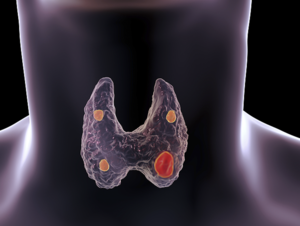
Primary hyperparathyroidism (pHPT) is the most common condition associated with the parathyroids. This disease is typically associated with high calcium levels. It is caused by one or more parathyroid tumors or parathyroid hyperplasia (enlargement of all four parathyroids).

Thank you for taking time to answer questions. I had 3 1/2 parathyroids removed six months ago. A month after surgery my PTH was 42 pg/ml, Vitamin D 25-OH was 38 ng/ml, but I’m confused what my calcium was as there were two results on the same day. One calcium level (in the metabolic panel) was 9.7 mg/dl and the other one that was listed with the PTH was 10.2 mg/dl. This confused me, but I felt better than I had in years so I wasn’t worried. However, it didn’t last long. The bone pain, fatigue and brain fog ( to name a few) returned and consistently have gotten worse. I have been taking calcium with vitamin D and magnesium as instructed after surgery. About 3 weeks ago I stopped taking it because the bone pain has gotten worse and I feared my calcium was high again. I am right back to feeling like I did pre surgery. I’m very disappointed and discouraged. My most recent lab results show that my calcium is 10.2 mg/dl. My vitamin D is 23 ng/ml and PTH is 17 pg/ml. I am 53 years old. What would cause my calcium to still be high and my other numbers to be so low? I’m so confused. I should probably mention I have an identical twin sister who has had a parathyroidectomy as well and still doesn’t feel well either, but she had a one sided surgery. Thank you for your time and I look forward to your response.
Thanks for writing. Your case is complex, and there are a few separate issues to unpack here. First, let's look at your most recent labs. Your calcium is 10.2 mg/dl, which is high for your age. Your PTH level is 17 pg/ml, which is low, in the suppressed range. This is a good sign, indicating that your remaining parathyroid tissue is responding appropriately to the high calcium. We want the ...
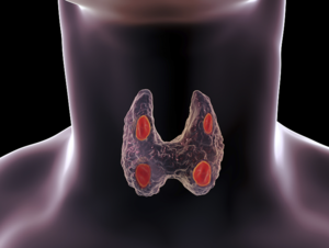
This question is regarding Direct Visualization of all 4 parathyroids versus radioguided parathyroidectomy. When directly visualizing the 4 parathyroids, is it easy to tell a normal gland versus a hyperactive gland? I'm sure it is easy to tell if one is grossly enlarged but if it is going abnormal (producing too much PTH) but not yet enlarged, can that be seen or can it only detected by the radioactivity? If 2 of the glands are abnormal or will become abnormal, do they usually become abnormal at the same time? The reason I ask this question is that I see you worked at the Norman Parathyroid Center who uses the radioguided method while you prefer the direct visualization method. Do you still run a sestamibi scan before each surgery but simply don't take a sample of each gland to test individually for radioactivity? Is it just a difference in your views on sampling via visual and being able to tell by size or texture or other feature rather than radioactivity and consequential exposure for patients? Since 20% have more than one gland hyperactive, I'm wondering how certain the visualization is or if in certain cases you do test. I see your method is different now from what you did at Norman Center and just wondered why you changed.
Thank you for your question. It touches on several important aspects of parathyroid surgery. I was at the Norman Parathyroid Center for six years, and was the Medical Director when I left to start my own practice. In starting my own center, I took a close look at our practices in Florida and critically evaluated each one. For the most part, the operation that I do here is the same as the one I ...